We talked about some rotator cuff exercises. And we will discuss a lot more. For now lets hop into some quick shoulder anatomy.
This will help you understand the shoulder, which will grow your ability to care for it.
Remember, your arm is your most important asset. Know it well!
The Shoulder – Anatomy
The shoulder is one of the most vital and essential body parts for conducting motions and movements. The shoulders are multi-directional and, due to their particular anatomy, they are capable of moving in all four directions – upwards, downwards, forwards and backwards – as well as being able to rotate in circular motions. [Obvious to most]
The basic anatomy of the shoulder will now be defined using illustrations for better understanding of the structure.
Shoulder Bones
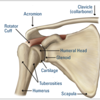
Figure 1 shows the basic structure of the shoulder. There are three main bones attached at a 90-degree angle to create the shoulder joint. These are as listed below.
- Humerus: the bone in the upper arm
- Scapula: the shoulder blade
- Clavicle: the collarbone
These bones are joined together to make two different joints. Along with each bone, there are attached muscles that provide extra protection and aid in movement.
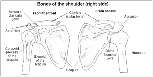
Figure 1 – Structure of the Shoulder
Shoulder Joints
The three bones of the shoulders make two joints, which are the gleno-humeral joint and the acromio-clavicular joint. Figure 2 shows, that the connection between the arm and the scapula (shoulder blade) make a gleno-humeral joint. The joint between the scapula and clavicle (collarbone) make the acromo-clavicular joint.
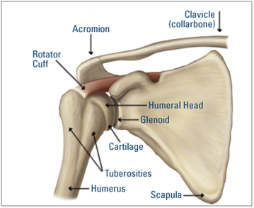
Figure 2 – Joint in Shoulder Muscles
Shoulder Muscles and Ligaments
The body is composed of four main elements, which collectively create the structure of the human skeleton. These are the bones, muscles, tendons and ligaments. A muscle is made up of tissues that contract and relax in accordance with nerve signals. A tendon connects muscles with bones and a ligament connects bones with bones.
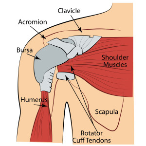
Many muscles hold the shoulder joint together. The most important of all is the rotator muscle and the acromio-clavicular ligaments, as well as the gleno-humeral joint capsule – as shown in Figure 3. The gleno-humeral joint capsule is a ball and socket joint that is formed by the humerus and scapula. This joint helps in the circular rotation of the arm, as well as its upward and downward movement. It also allows the arm to move freely in the socket.
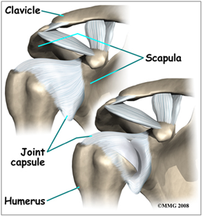
Figure 3 – Gleno-humeral Joint Capsule
The other surrounding muscles in the area also support the movement of the shoulder. They are the deltoid shoulder muscle, pectoralis major (chest muscles), teres major, lattissimus dorsi, biceps and triceps. The biceps allow the elbow to bend instantly while the other six muscles help with rotator cuff injuries and conditions. The deltoid muscles constitute of the anterior fiber, posterior fiber and middle fiber. The deltoid anterior fiber flexes the shoulder and horizontally abducts it. The posterior fiber deltoid helps in the extension of the muscle and abducts the shoulder horizontally. Finally, the middle deltoid fiber abducts the shoulder.
Get it? Good!
Next Post:
Next time we will get into the roator cuff anatomy and why its important. So stay tuned.
Thanks for reading, share with someone important.
-Zach
PS: Do you want a free book of 18 rotator cuff exercises zach does everyday? click here


Leave A Reply (No comments so far)
No comments yet